- Home
- Leg Alignment Surgery in Case of Joint Overload due to Misalignment
Leg Alignment Surgery in Case of Joint Overload due to Misalignment
SYMPTOMS
You have felt discomfort in your knee joint after weight-bearing exertion for some time. The orthopedic picture-book leg is straight and has a slight X-shape. With this form, the loads are distributed approximately evenly on the inside and outside. Many athletes have O-shaped (bow) legs, and thus the pressure load runs on the inside. Everything goes well until the balance of this biological system is disturbed: an injury to the anterior cruciate ligament (ACL), a medial meniscus lesion, or cartilage injury are sufficient, and the knee is no longer able to recover on its own.
A balanced leg axis that keeps the pressure away from the damaged side is the basis of knee-preservative therapy. Attempts to repair an overloaded inner- side, with meniscus or cartilage therapy, have no chance when the foundation is lacking.
What you feel: During or after major exertion, pain on the inside that is difficult to grasp and describe and possibly slight swelling of the knee. The resilience of your knee joint is limited and your quality of life is significantly restricted.
EXAMINATION
We often see the typical bow leg configuration during the examination. If your gait shows an increased bending of the leg in an O-form, it is a critical situation. Then not only the inside is under pressure, but the ligaments on the outside are under tension, and the problem increases. Swelling, tenderness, which are the typical signs of inner side overloading, are confirmed on X-ray, MRI and possibly SPECT/ CT images.
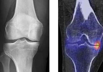
LEGENDE
1 – X-ray right knee front view with narrowing of the joint space on the inside
2 – SPECT/CT right knee shows overload on the inside
TREATMENT
Physiotherapy, medication or splints can be used to try to relieve the symptoms. These cannot solve the basic bio-mechanical problem. However, these remedies can be helpful to bridge the gap until the operation.
SURGICAL PROCEDURE
The leg axis can only be corrected by changing the shape of the bone. Most often, this requires a change in the shape of the lower leg, which is achieved by an intervention directly below the knee joint. We call this procedure an osteotomy. The location and extent of the correction is precisely planned before the operation. We often start the operation with knee mirroring (arthroscopy) to treat existing problems in the joint.
Next, the region on the leg below the knee joint is exposed, and we perform the osteotomy under fluoroscopic guidance. We stabilize the newly obtained bony shape with a plate and screws so that the bone can heal in the desired shape. Duration of the operation: 1 hour.
This decription concentrates on the bow leg configuration, as this is more common. The exact same bio-mechanical problem occurs when you have an X-shaped leg (Knocked-knee) and the outside of your knee is worn out. Duration of the operation: 1.5 hours.
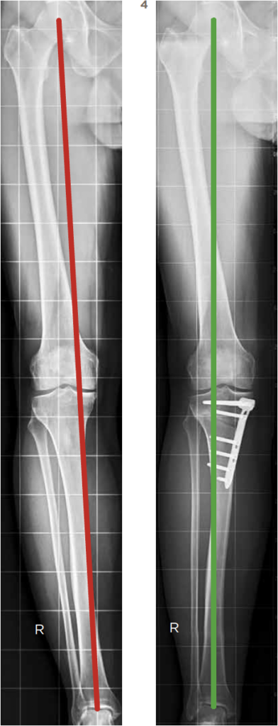
LEGEND
3 – X-ray full right leg before correction: bow leg
4 – X-ray full right leg after correction: straight leg
RISKS
You will be treated by experienced surgeons. And yet it is like flying: no surgery is without risk. The risks of this operation can be summarized as follows:
Risk of infection: ~ 1%
Likelihood that you will require a blood transfusion: less than 1%
Damage to relevant blood vessels: 1 – 2%
Damage to relevant nerves: 1 – 2%
Thrombosis/Embolism: 2 – 5%
Delayed bone healing: considerably less than 1%, higher in smokers
Breakage of the osteosynthesis materials: considerably less than 1%, higher in smokers
HOSPITAL STAY
After the operation, your leg is placed in an immovable cloth splint, as we want to give the leg a fair amount of rest at the beginning. As soon as you can get up well and have yourself and your leg under control again, we will remove this splint. As a rule, you stay hospitalized for 4–5 days.
DISCHARGE
The stitches can be removed by your family doctor about 12 – 14 days after the operation. We will routinely see you for check-ups in the out-patient clinic after 6 weeks, and 3 and 6 months (with X-rays). Usually you should be fine after 6 months and the bone will have healed to the point that we can plan the metal removal at a convenient time.
WEIGHT-BEARING, what happens and when?
Transition to walking without crutches: after 6 weeks
Good, limp-free walking: after 3 months
Sport, depending on the type of sport: after 3 – 12 months
pdf for download:Leg Alignment Surgery